Fig. 3.

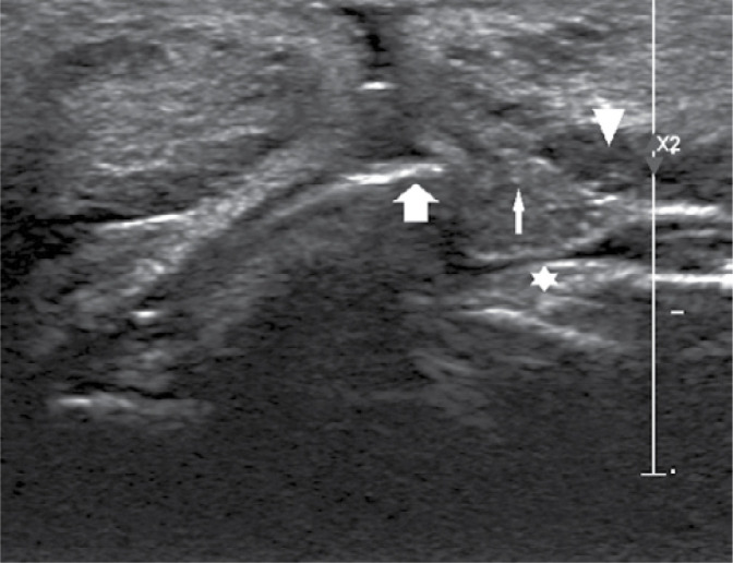
Ultrasound image (Philips Epiq 5) of a hyaluronic acid deposit (arrow head) in the lower lip pushing out a labial gland (thin arrow) catching on the upper incisor (bold arrow); maxillary mucosa (asterisk)

Ultrasound image (Philips Epiq 5) of a hyaluronic acid deposit (arrow head) in the lower lip pushing out a labial gland (thin arrow) catching on the upper incisor (bold arrow); maxillary mucosa (asterisk)