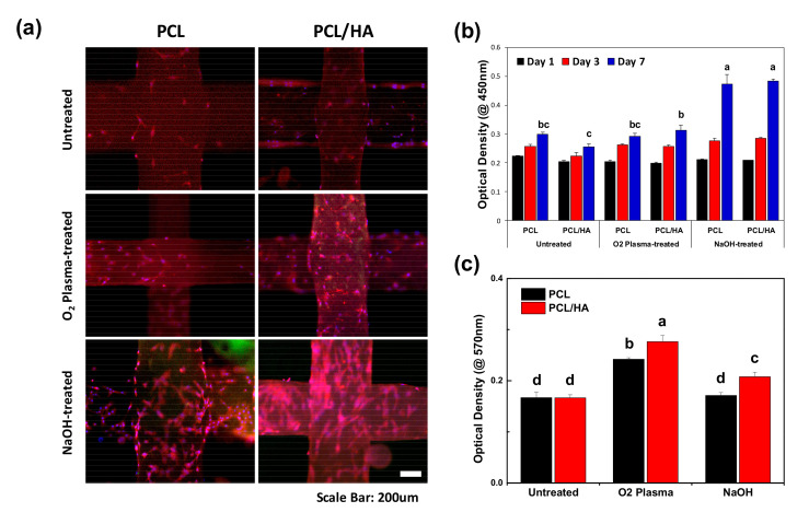
Figure 3.
In vitro study of hDPSCs on the scaffolds. (a) Fluorescence microscopy image of the hDPSCs attached on the scaffolds on day 3. Both O2 plasma and NaOH treatments enhanced cell attachment of the scaffolds. (b) Cell proliferation on days 1, 3, and 7 using a WST-1 assay (n = 7, ANOVA, Duncan’s multiple range test, p < 0.05). At day 7, the NaOH-treated scaffolds exhibited significantly enhanced cell proliferation. Same letters indicate that there is no significant difference between samples. (c) Measurement of protein adsorption on the scaffolds after 24 h of incubation with 1% bovine serum albumin (BSA) solution (n = 5, ANOVA, Duncan’s multiple range test, p < 0.05). O2 plasma treatment greatly promoted protein adsorption ability of the scaffolds. Same letters indicate that there is no significant difference between samples.

