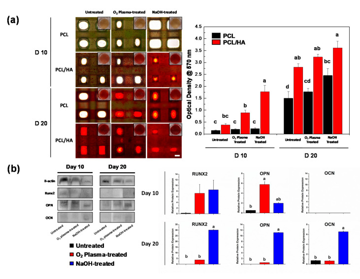
Figure 4.
Osteogenic differentiation of hDPSCs on the scaffolds (a) Alizarin Red S staining of the hDPSCs cultured on the scaffolds on days 10 and 20. The quantitative data was obtained using de-staining (scale bar: 500 μm). The NaOH-treated PCL/HA group exhibited enhanced calcium deposition. Same letters indicate that there is no significant difference between samples. (b) The expression of osteogenic proteins on the PCL/HA scaffolds (RUNX2, OPN, and OCN) was determined by Western blotting on days 10 and 20. Quantitative results showed significantly enhanced RUNX2, OPN, and OCN expression on the NaOH-treated PCL/HA scaffold. (n = 3, ANOVA, Duncan’s multiple range test, p < 0.05). Same letters indicate that there is no significant difference between samples.

