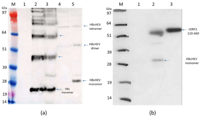
Figure 3.
Western blot analysis of HBcAg and HBcHEV ORF2 551–607. (a) Western blot with mouse anti-HBcAg monoclonal antibody (10E11, Abcam, UK). M, marker; 1, pEAQ-HT empty vector; 2, HBcAg supernatant (SN) after extraction in 3x volume extraction buffer and 13k rpm; 3, HBcAg pellet; 4, HBcHEV ORF2 551–607 SN; 5, HBcHEV ORF2 551–607 pellet; (b) Western blot with polyclonal anti-HEV IgG swine serum. M, marker; 1, pEAQ-HT empty vector; 2, HBcHEV ORF2 551–607 crude extract; 3, positive control rHEV 110–660. Arrows indicate monomer, dimer, and tetramer. SeeBlue Plus2 Pre-stained Protein Standard was used as a molecular marker.

