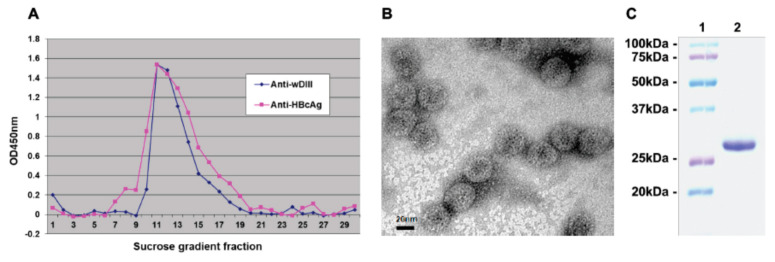
Figure 3.
VLP assembly of plant-produced HBcAg-wDIII. Protein extract from HBcAg-wDIII-expressing leaves was subjected to a 10–60% sucrose gradient centrifugation. (A) ELISA of sucrose gradient fractions. An antibody against wDIII or HbcAg was used to detect the wDIII or HbcAg moiety in HBcAg-wDIII, respectively. (B) Electron microscopy of HBcAg-wDIII from peak fractions of the sucrose gradient. Bar = 20 nm. One representative field is shown. (C) SDS-PAGE analysis. Lane 1: molecular weight marker; lane 2: HBcAg-wDIII from peak fractions of the sucrose gradient.

