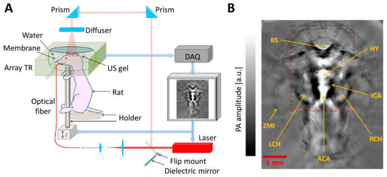
Figure 4.
Schematic illustration and representative images of a PACT system. (A) Typical configurations of PACT. Array transducer is used to acquire PA images. (B) Representative PACT images of mouse brain in vivo. PA, photoacoustic; PACT, photoacoustic computed tomography; TR, ultrasound transducer; US, ultrasound; DAQ, data acquisition module; BS, brain stem; HY, hypothalamus; ICA, internal carotid artery; LCH, left cerebral hemisphere; RCH, right cerebral hemisphere; ACA, anterior cerebral artery; ZMI, zygomatic muscle interface. The images are reproduced with permission from [42].

