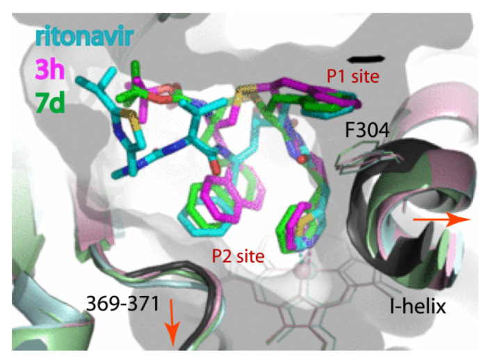
Figure 9.
Relative orientation of the best rationally designed inhibitors, 3h and 7d, and ritonavir (3NXU structure; molecule A). The respective structures are coloured in pink, light green and light cyan. Structure of water bound CYP3A4 (5VCC) is rendered in black. Displacement of the I-helix and the 369–371 fragment triggered by the inhibitor binding is indicated by red arrows.

