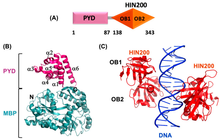
Figure 3.
Structural details of AIM2. (A) Schematic representation of AIM2 domain organization; (B) crystal structure of AIM2PYD with Maltose-binding protein (MBP) fusion tag (PDB ID: 3VD8) [161]; (C) crystal structure of AIM2HIN in complex with dsDNA (PDB ID: 3RN2) [162]. The ribbon diagrams are generated with PyMOL molecular graphics software.

