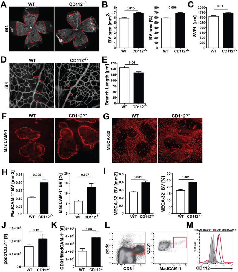
Figure 3.
CD112 deficiency increases blood vessel coverage in the retina and spleen in mice. (A–E) Whole mounts from postnatal 6 (P6) retinas were stained with isolectin B4 (iB4) to visualize the blood vessels of the superficial vascular plexus, followed by quantification of vascular parameters. (A) Representative confocal images of iB4 stained P6 retinal whole mounts of CD112−/− and Wild type (WT) pups. 4× objective 1.0 zoom. Image-based morphometric analysis showing (B) the iB4+ area (left) and percent covered by blood vessels (right) and (C) the superficial vascular plexus length (SVPL). Scale bar: 500 µm. Data from one out of 3 similar experiments (n = 4–6 mice/group). (D,E) Upon vessel maturation, vessels undergo pruning, which can be quantified by analysing the blood vessel density. (D) Representative images of P6 retinas of WT and CD112−/− stained with iB4. 20× objective, 1.0 zoom, scale bar: 80 µm. Analysis of (E) the branch length formed in the peri-arterial space in CD112−/− mice. Data from one experiment are shown (n= 4–6 mice/group). (F–M) Immunofluorescence and flow cytometry analysis of spleen single-cell suspension. (F–I) Sections sizes of 40 μm of optimum cutting temperature (OCT)-frozen spleens from CD112−/− and WT mice were stained for MadCAM-1 (F) and MECA-32 (G). Representative pictures are shown. Scale bar: 200 µm. (H,I) Quantification of the MadCAM-1+ (H) and MECA-32+ (I) area in spleen of CD112−/− mice; n = 5 mice/group. (J,K) Quantification of the number of (J) podo−CD31+ and (K) CD31+MadCAM-1+ BECs present in the spleen of WT and CD112−/− mice. Pooled data from three experiments are shown. (L) Gating strategy of spleen single-cell suspensions. BECs were gated on CD45−podo−CD31+ (left), MadCAM-1+ CD31+ (right). (M) Representative histogram of several independent experiments showing CD112 expression by BECs in the spleen. Blue: CD31+, red: CD31+MadCAM-1+, grey: isotype controls.

