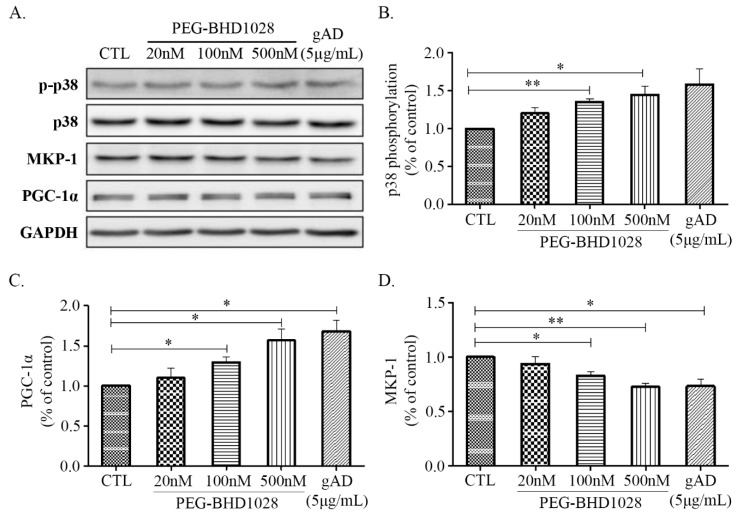
Figure 5.
The regulatory effects of PEG-BHD1028 on mitochondrial biogenesis in C2C12 myotubes. (A) The activation of p38 MAPK, PGC-1α, and MKP-1 following the treatment with PEG-BHD1028 was observed by western blot. (B) The change in the ratio of the phosphorylation band density of p38 MAPK to control MAPK was quantified. (C,D) The quantification of PGC-1α and MKP-1 proteins was represented as a ratio to GAPDH. Results are presented as means ± SEM (n = 3). ** p ≤ 0.01, * p ≤ 0.05 vs. CTL (Negative control).

