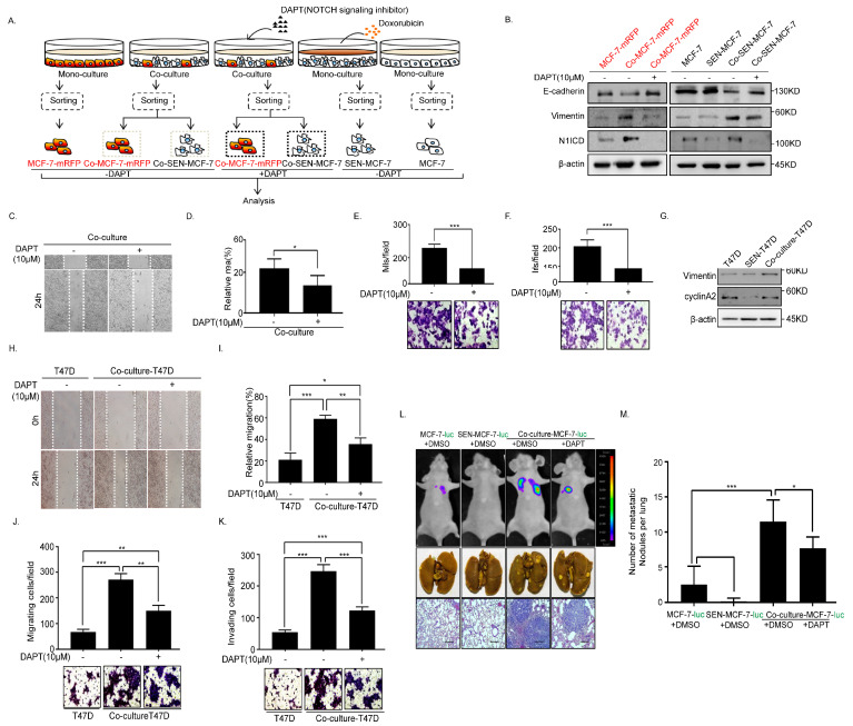
Figure 4.
N-[(3,5-Difluorophenyl)acetyl]-l-alanyl-2-phenyl]glycine-1,1-dimethylethyl ester (DAPT) inhibits EMT and metastasis of the senescent and adjacent non-senescent breast cancer cells. (A) Scheme showing the experimental setup. Flow cytometry analysis of collected breast cancer cells in co-cultures or monocultures treated with or without DAPT (10 μM). (B) Immunoblot analysis of protein expression of active Notch1 intracellular domain (N1ICD), E-cadherin, and Vimentin in MCF-7-mRFP and MCF-7 cells treated with or without DAPT. (C,D) Wound-healing assay of MCF-7 cells (MCF-7-mRFP and SEN-MCF-7) treated with or without DAPT. (C) Representative micrographs of the wound-healing assay are shown. (D) Data represent the relative migration ratio of cells per field (error bars indicate mean SD, n = 3 experimental replicates, * p < 0.05). (E) Migration assay of MCF-7 cells (MCF-7-mRFP and SEN-MCF-7) treated with or without DAPT. Representative micrographs of migrated cells are shown. Data represent the number of cells derived from mean cell counts of five fields (error bars indicate mean SD, n = 3 experimental replicates, *** p < 0.001). (F) Invasion assay of MCF-7 cells (MCF-7-mRFP and SEN-MCF-7) with indicated treatments. Representative micrographs of the migrated cells are shown. Data represent the number of cells derived from mean cell counts of five fields (error bars indicate mean SD, n = 3 experimental replicates, * p < 0.05, ** p < 0.01, *** p < 0.001). (G) Immunoblot analysis of protein expression of E-cadherin, Vimentin, N1ICD, and cyclinA2 in T47D, SEN-T47D and Co-culture-T47D (which contains T47D and SEN-T47D). (H,I) Wound-healing assay of T47D subjected to the indicated treatments. (H) Representative micrographs of the wound-healing assay are shown. (I) Data represent the relative migration ratio of cells per field (error bars indicate mean SD, n = 3 experimental replicates, * p < 0.05, ** p < 0.01, *** p < 0.001). (J) Migration assays of T47D cells with the indicated treatments. Representative micrographs of migrated cells are shown. Data represent the number of cells derived from mean cell counts of five fields (Error bars indicate mean SD, n = 3 experimental replicates, ** p < 0.01, *** p < 0.001). (K) Invasion assay of T47D cells with the indicated treatments. Representative micrographs of migrated cells are shown. Data represent the number of cells derived from mean cell counts of five fields (error bars indicate mean SD, n = 3 experimental replicates, *** p < 0.001). (L) Lung metastatic nodules were confirmed by hematoxylin and eosin staining. Scale bars, 250μm. (M) The number of visible surface metastatic lesions in lungs was counted (error bars indicate mean SD, n = 6 mice for each group, * p < 0.05, *** p < 0.001).

