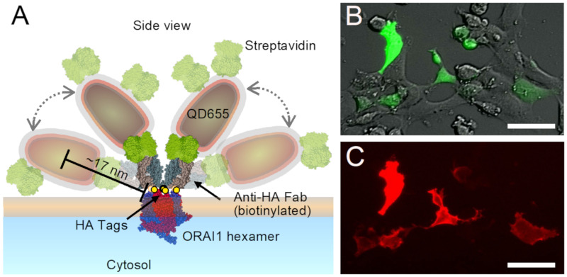
Figure 1.
Labeling of ORAI1 proteins in the plasma membrane of HEK cells with quantum dots (QDs). (A) Schematic representation of QD-labeling of the ORAI1 Ca2+ channel in the plasma membrane. Labels consisted of a biotinylated anti hemagglutinin (HA) antigen-binding fragment (Fab) that bound to the HA-tags (yellow spheres) at the extracellular region of each subunit of the ORAI1-HA channel, shown in hexameric conformation (blue and red). The bound Fab was labeled using streptavidin (green) conjugated fluorescent QDs with an electron-dense core (dark gray), and an electron transparent polymer coating shell (red/grey). Several possible spatial positions are shown of two exemplary bound QDs, the positions showing half-transparent labels represent a maximally extended configuration. The representation of all involved molecules reveals a maximum distance between the center of a bound QD and the ORAI1 hexamer of 17 nm. All protein structures were derived from the Protein Data Base, sizes of QDs were measured elsewhere [9], and all structures are drawn to scale. (B) Overlay of direct interference contrast light microscopy and fluorescence microscopy of green fluorescent protein (GFP) intracellularly co-expressed with the same ratio as ORAI1. (C) Fluorescence images of QD-labeled ORAI1 at rest showing overlap with the cells expressing ORAI1 visible from GFP in (B). Scale bars = 50 μm.

