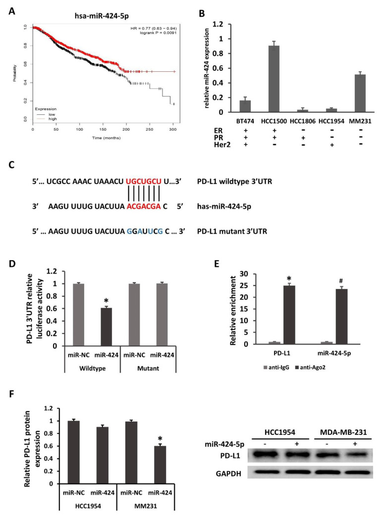
Figure 2.
PD-L1 is directly regulated by miR-424-5p. (A) Kaplan–Meier overall survival analysis (from 1262 cases of breast cancer, kmplot.com, ID in KMPLOT: hsa-miR-424) revealed that patients with high miR-424-5p expression exhibited longer overall survival times. p = 0.0091. miR, microRNA; HR, hazard ratio. (B) miR-424-5p expression in several breast cancer lines, compared with the normal breast epithelial MCF-10A cell line. (C) PD-L1 is a potential target of miR-424. The binding sites in the 3′UTR of PD-L1 are shown. (D) Luciferase activity from cotransfection of wild-type (WT) and mutant (Mut) PD-L1 regions with miR-424 or negative control (miR-NC) in HEK293T cells. (E) RNA from MM231 cells precipitated with anti-Ago2 or nonspecific rabbit IgG were analyzed via qRT-PCR assay. Relative quantification for PD-L1 transcript was performed using GAPDH mRNA as a reference gene. The relative expression of PD-L1 protein in HCC1954 and MM231 (F) were determined by Western blotting 48 h after transfection. Relative quantification for PD-L1 protein was performed using GAPDH protein as a reference protein. * p < 0.05, # p < 0.05.

