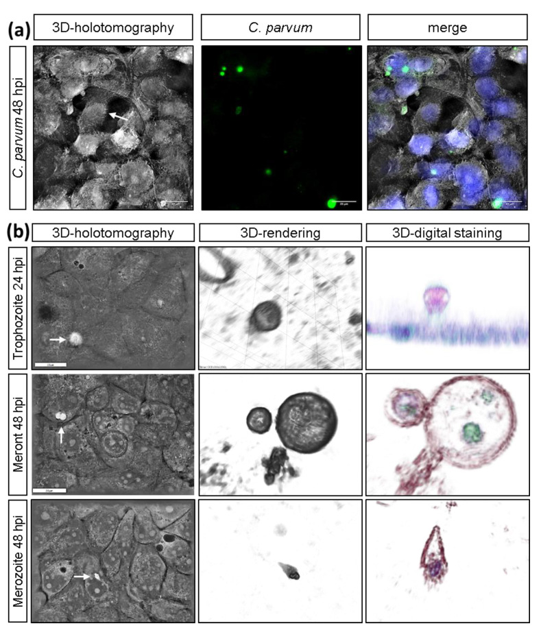
Figure 1.
3D-holotomographic illustration of C. parvum-infected HCT-8. (a) C. parvum-infected HCT- 8 cells were stained by biotinylated VVL and Hoechst 33258 at 24 and 48 h p. i. (n = 3): (a) 3D-holotomographic images were obtained by using 3D Cell Explorer microscope (Nanolive) at 60× magnification (λ = 520 nm, sample exposure 0.2 mW/mm2) and a depth of field of 30 µm. (b) Holotomography of C. parvum-infected HCT-8 and identification of trophozoite, meront and merozoite stages (Scale bar = 20 µm).

