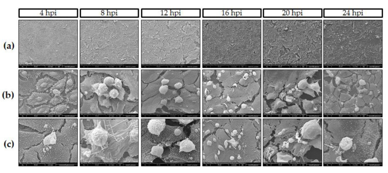
Figure 2.
C. parvum development in HCT-8 cells under hyperoxic (21% O2) conditions (n = 6). SEM-based illustration of C. parvum-infected HCT-8 evidenced rapid C. parvum development and infection kinetics: (a) Overviews at different time points post infection (4–24 h p. i.). (b,c) Closer views of parasite stages-host cell interactions, i.e., column at 16 h p. i. shows meront-infected cells, row b evidenced hole-like damage on host cell surface induced by merozoites release at the same time point (white arrow). Row c (4 h p. i.) reveals early induction of villi-like structures on surface of infected cells (black arrows).

