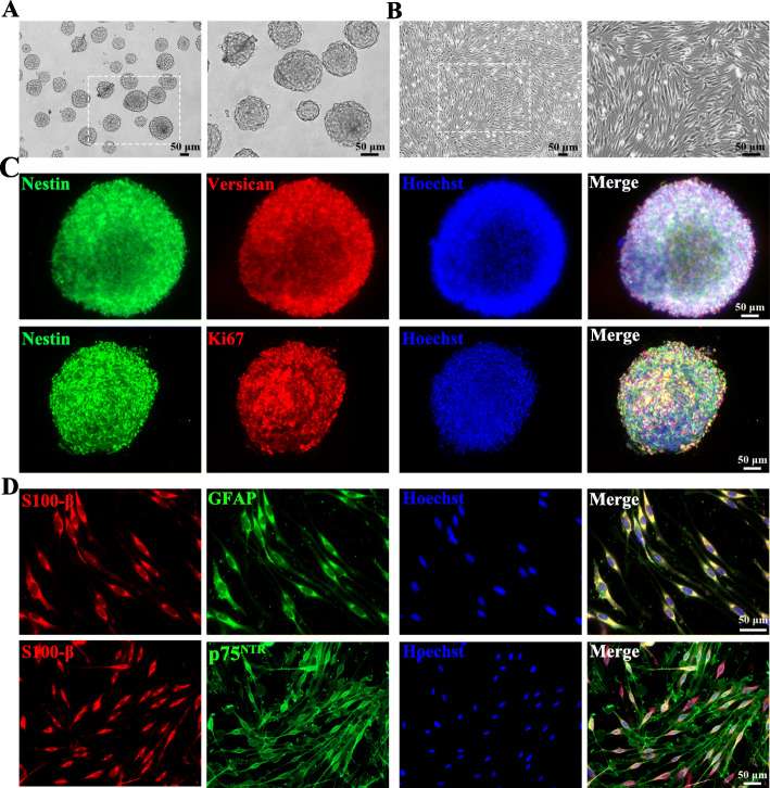
Fig. 1.
Characterization of SKPs and SKP-SCs. a SKPs in floating culture showed spherical colonies. b SKP-derived SCs showed spindle-like shape with side-by-side alignment. c Immunofluorescence staining of SKP spheres showed positive expression of intermediate filament Nestin (green) and chondroitin sulfate proteoglycan Versican (red) with proliferation marker Ki67 (red) and Hoechst (blue) labeled cell nuclei. d SKP-derived SCs showed positive expression of SC markers S100 (red), GFAP (green), and p75NTR (green), and cell nuclei were labeled with Hoechst (blue). Scale bar, 50 μm

