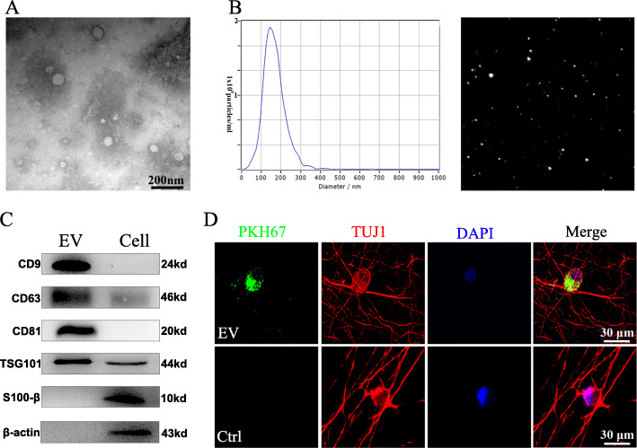
Fig. 2.
Characterization of SKP-SC-derived EVs. a Representative TEM image of SKP-SC-EVs presenting a typical cup-like concavity shape. Scale bar, 200 nm. b Representative traces from nanoparticle tracking analysis for SKP-SC-EVs. c Western blots showing the positive expression of exosomal markers CD9, CD63, CD81, and TSG101 in EVs, and β-actin and S100β were used as control markers in SKP-SCs. d PKH67 labeled EVs (green) were showing within the cytoplasm and axon of TUJ1 positive sensory neurons (red), and nuclei were labeled with DAPI (blue). Scale bar, 30 μm

