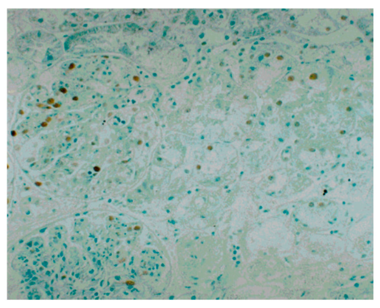
Figure 5.
Immunoperoxidase staining for proliferating cell nuclear antigen (PCNA) and counterstaining with methyl green in a renal biopsy tissue from a PSAGN patient (original magnification, ×100) [23]. Prominent expression of PCNA in the glomerulus and tubular epithelial cells was observed. The distribution of PCNA-positive cells was variable; i.e., there was a considerable difference in the number of PCNA-positive cells between each glomerulus and between different parts of the tubules.

