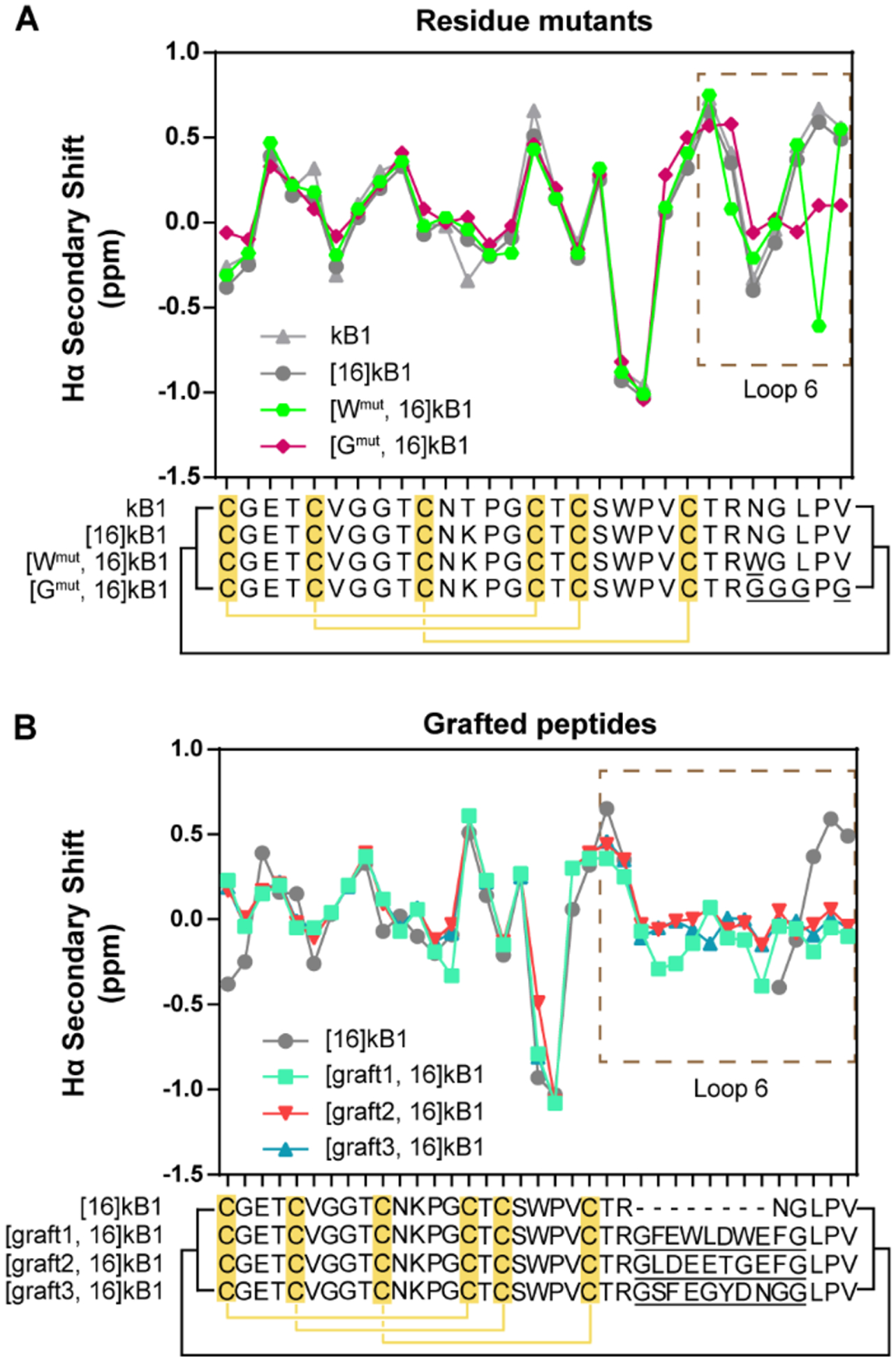
Figure 4.

Structural characterization of cyclotide analogues using NMR. (A) Hα chemical shift analysis of kB1 analogues with mutations in loop 6 (residue mutants). (B) Hα chemical shift analysis of kB1 analogues with epitopes grafted into loop 6 (grafted peptides). Each point in the line graph presents the Hα secondary shift of an individual residue, and the sequence alignment of the peptides is shown below, with mutated residues in loop 6 underlined. The disulfide connectivity is indicated by yellow lines and the cyclic backbone by black lines. The region of kB1 with mutations and grafted epitopes (loop 6) is highlighted in a dashed line box.
