
Figure 5. Expression of LOX-1 protein is elevated in response to ox-LDL and within atherosclerotic lesions.
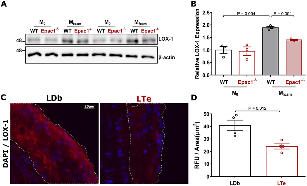
(A) Representative immunoblot of LOX-1 and β-actin expression in WT and Epac1−/− differentiated M0 and Mfoam BMDMs treated with vehicle or ox-LDL (40 μg/mL, 48 h), respectively. (B) Quantification of relative LOX-1 protein expression (N = 3 independent mice per genotype). (C) Representative images of LOX1 immunofluorescence staining in lesions from sagittal sections of the aortic arch. (D) Quantification of immunofluorescence signal per lesion area in LDb and LTe lesions, where a minimum of two independent fields of view were processed for each animal (N = 4 mice per genotype). Data are presented as mean ± SEM.