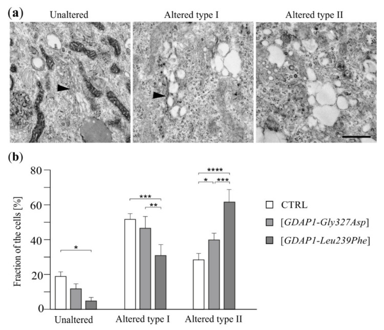
Figure 3.
Expression of mutant GDAP1 variants affects the morphology of the Golgi in HeLa cells. (a) Transmission electron microscope analysis of the Golgi in non-transfected HeLa cells (CTRL) or in HeLa cells transfected with a vector carrying GDAP1-Gly327Asp or GDAP1-Leu239Phe alleles. Electron micrographs of representative examples of Golgi structures observed in HeLa cells. Black arrowheads indicate the cisternae of the Golgi. Scale bar 750 nm. (b) Fraction of cells [as percentage of counted cells] with types of Golgi ultrastructure indicated. Statistical analysis was performed on data from three independent experiments using one-way ANOVA and Bonferroni’s correction: * p < 0.05, ** p < 0.01, *** p < 0.001, **** p < 0.0001.

