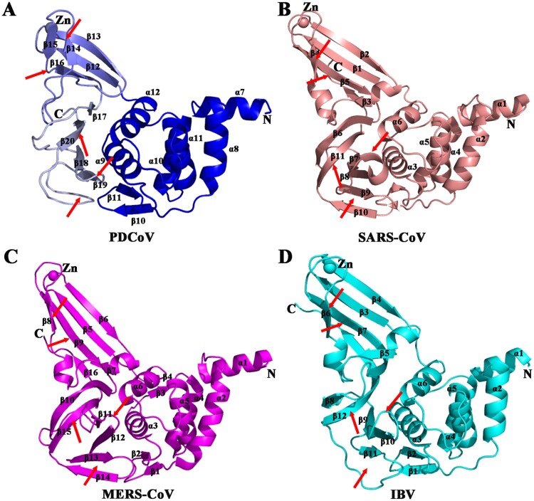
Figure 3.
Structural comparison of PLPs among four CoVs. (A) Detailed structure of PDCoV PLpro from δ-CoV (blue). (B) Structure of SARS-CoV PLpro from β-CoV (PDB ID: 2FE8, salmon[27]). (C) Structure of MERS-CoV PLpro from β-CoV (PDB ID: 4RNA, magenta [60]). (D) Structure of IBV PLpro from γ-CoV (PDB ID: 4X2Z, cyan [24]). From A to D, the secondary structures (helices, strands, and loops) are marked; Zn2+ ions are shown as spheres. Regions that show significant differences among genera are indicated by arrows.

