
Figure 1.
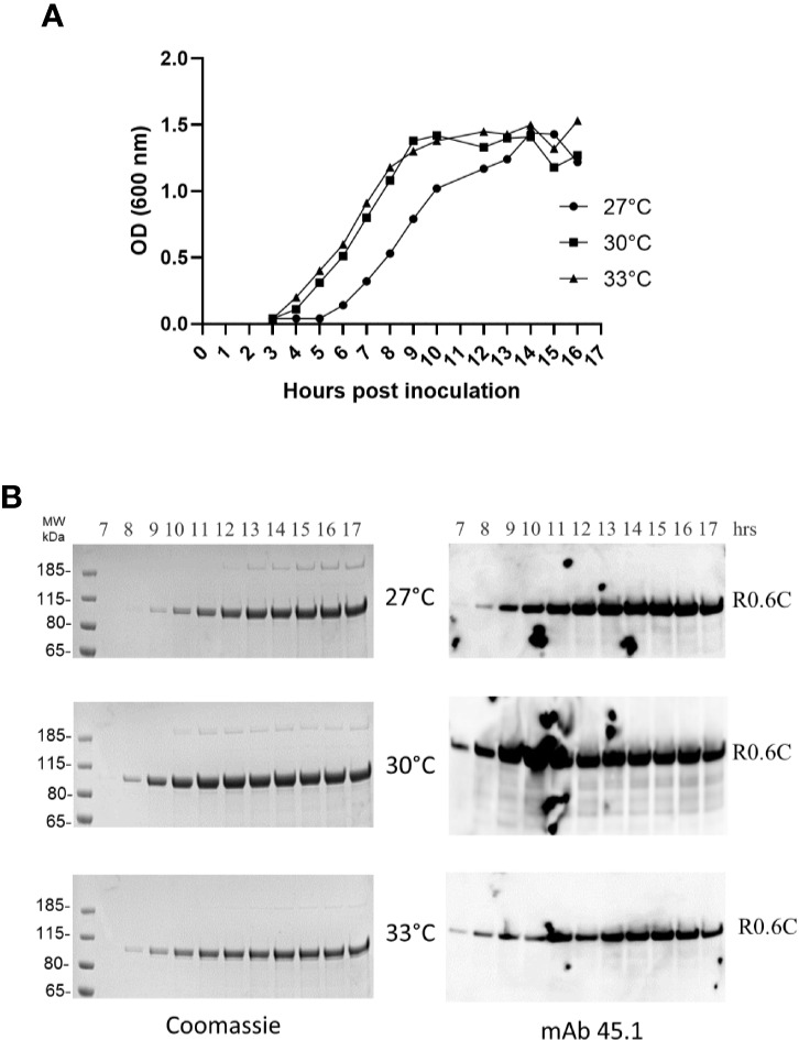
Time course analysis of R0.6C expression in L.lactis at different temperatures. (A) Optical density measurements at 600 nm from 3 to 17 h of bacterial growth at 27, 30, and 33°C. (B) Samples collected from 7 to 17 h were assessed by Coomassie and immune blotting analysis. Samples were analysed without a reducing agent. Left panel, Coomassie blue-stained 4%–12% polyacrylamide gel. Right panel, immune blotting analysis of the same gel shown to the left using the conformational reduction-sensitive mAb45.1 as primary antibody.