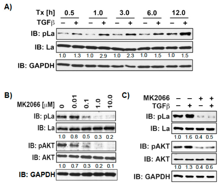
Figure 8.
TGFβ treatment increases phosphorylation of La at T389. (A) SCC22A cells cultured in serum-free medium were treated with TGFβ [5 ng/mL] or vehicle [1 mg/mL BSA in 4 mM HCl] for increasing time intervals from 0.5 to 12h. (B) SCC22A cells cultured in serum-free medium were treated with increasing MK2066 concentrations [0–10 µM] for 3 h followed by insulin treatment [1 µM] for 15 min. (C) SCC22A cells cultured in serum-free medium were treated with MK2066 [10 µM] or vehicle for 3 h followed by treatment with TGFβ [5 ng/mL] or vehicle [1 mg/mL BSA in 4 mM HCl] for 1 h. IB: immunoblot. Numbers below bands show the phosphorylation status of La or AKT by calculation the ratio of pLaT389/La or pAKTS473/AKT. The untreated control was set to 1 in relation to all other samples. GAPDH served as loading control.

