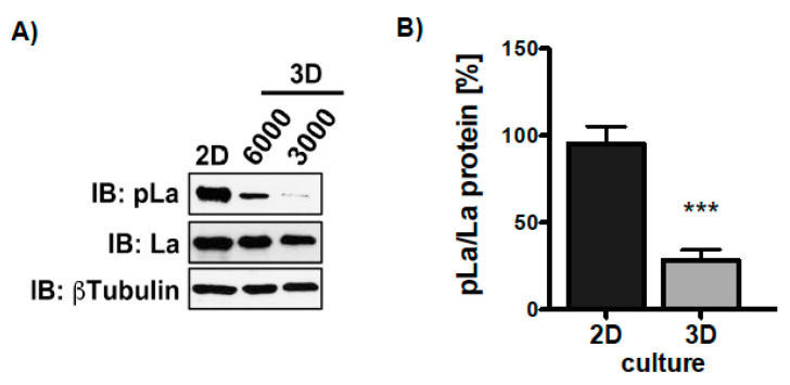
Figure 9.
More phosphorylation of La at T389 in 2D compared to 3D cultures. (A) Immunoblot (IB) of SCC22A cells 2D-cultured compared to SCC22A cells (3000 or 6000 cells plated) cultured on ultra-low attachment plates (3D sphere culture) in serum-free medium plus TGFβ [5 ng/mL]. (B) Quantitative analysis of the pLaT389/La protein ratio in 2D versus 3D cultures of SCC22A cells. The βTubulin served as loading control (p value < 0.001 (three asterisks), n = 3).

