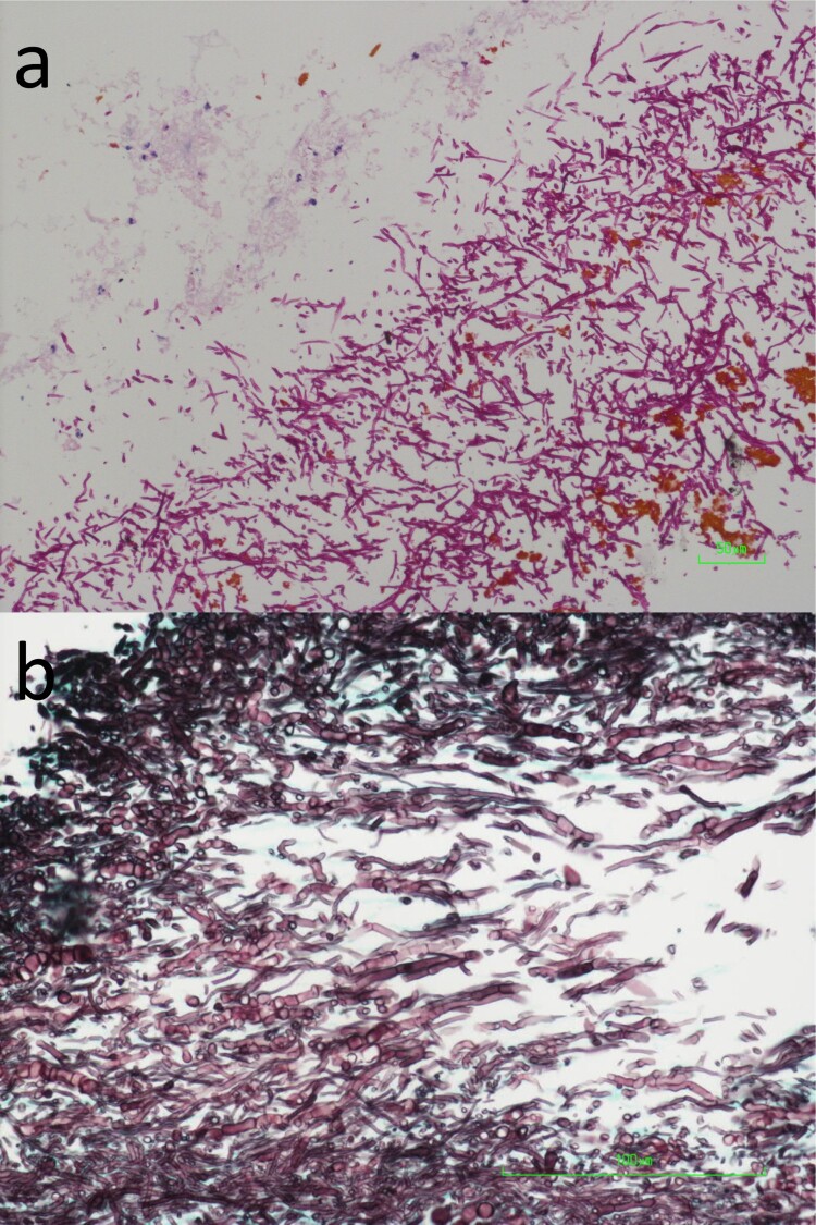
Figure 2.
Photomicrographs of the biopsied liver abscess wall tissue. (a) Abundant fungal hyphae, a small amount of necrotic tissue, brown bile pigments as well as a few acute inflammatory cells were observed (Periodic acid-Schiff staining, original magnification 200×). (b) The fungal hyphae were highly septate (Grocott methenamine-silver staining, original magnification 400×).

