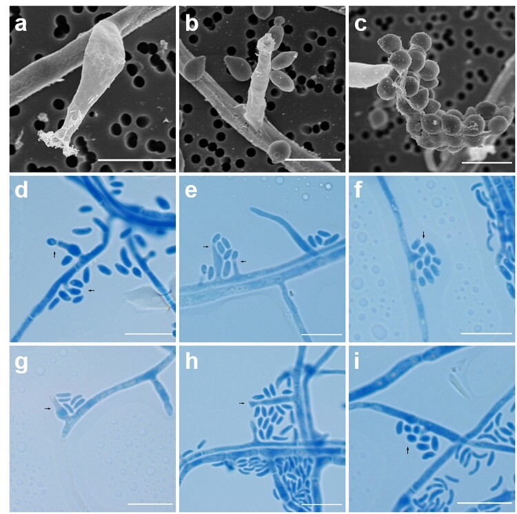
Figure 4.
Microscopic features of Pleurostoma hongkongense HKU44T. Arrows indicate notable structures. (a–c) Scanning electron microscopy photographs of notable phialide and conidia features. Scale bars = 5 μm. (d–i) Bright-field microscopy of corresponding features using a slide culture preparation. Slides were prepared via wet mount and stained with lactophenol cotton blue (original magnification 1000×). Scale bars = 10 μm.

