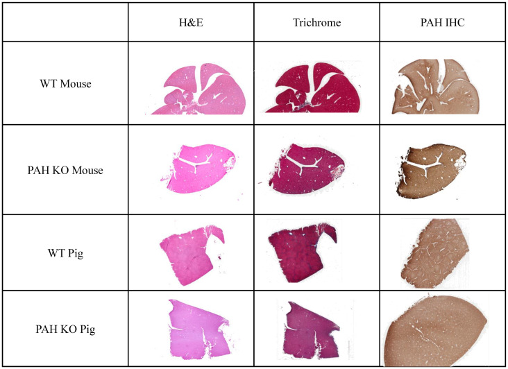
Fig 4. Histology of WT and Pah KO livers in mouse and pig.
Standard H&E and Masson’s trichrome sections show no appreciable difference between WT and Pahenu2/enu2 mice or WT and PAHR408W/R408W pigs. Similarly, immunohistochemistry showed similar levels and ubiquitous distribution of expression of the wild type and mutant protein in both models/species.

