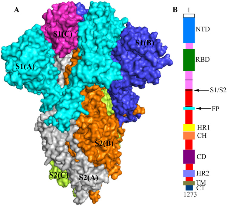
Fig 1.
(A) The structures of the S protein trimer (electrostatic potential surface area (PDB ID: 6vxx)),S1(A), S2(A), S1(B), S2(B), S1(C) and S2(C) are shown in cyan, gray, blue, orange, magenta, and yellow-green, respectively. (B) Schematic of the SARS-CoV-2 S protein primary structure, colored by domain.

