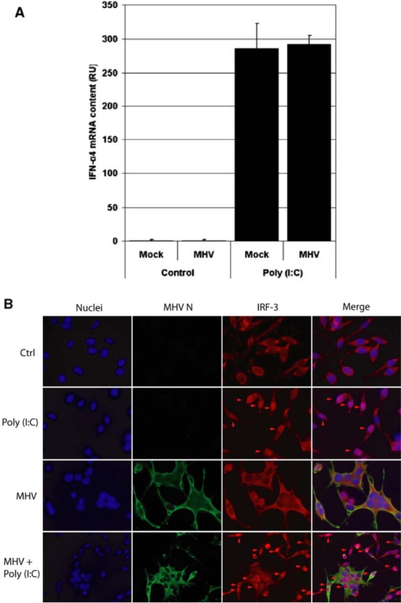
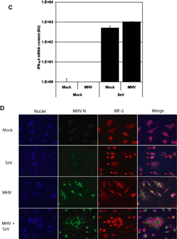
Fig. 2.
L-AC2 cells were infected with MHV at MOI 10. At 1 h p.i., cells were infected with 4 µg poly(I:C). At 8 h p.i. total RNA was isolated and reverse transcribed using random hexamers. (A) IFN-α4 mRNA concentration was determined using specific RT-qPCR and (B) endogenous IRF3 localization and MHV nucleocapsid protein were detected in IFA with specific poly- and monoclonal antibodies, respectively. Nuclear IRF3 is indicated with arrows. Subsequently, L-ACE2 cells were seeded on coverslips in 35-mm wells and infected with SeV for 30 min. Next, cells were infected with MHV at MOI 5. At 8.5 h.p.i. cells on coverslips were fixed in 3% paraformaldehyde. From the remainder of the cells, total RNA was isolated and reverse transcribed using random hexamers. (C) IFN-α4 mRNA concentration was determined using specific RT-qPCR and (D) endogenous IRF3 and MHV nucleocapsid protein were detected in IFA with specific poly- and monoclonal antibodies, respectively. Nuclear IRF3 is indicated with arrows. Figures modified from Ref. [8] and used with permission.


