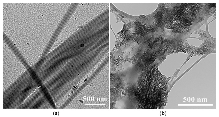
Figure 1.
Typical transmission electron microscopy (TEM) images of (a) highly oriented intrafibrillar (showing clear D-banding) and (b) extrafibrillar mineralized collagen fibrils. Images are modified from [24] with permission. Copyright @ John Wiley & Sons, Inc., Hoboken, NJ, USA.

