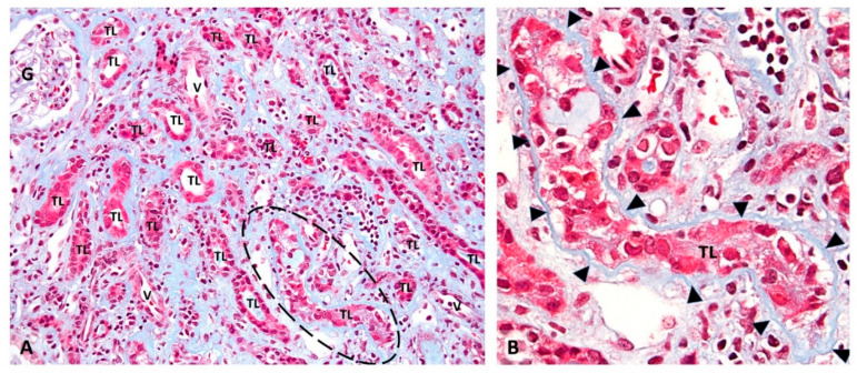
Figure 1.
Renal injury in “definitive” PyVN. Renal biopsy: (A) low-power view of a representative case of PyVN demonstrating many tubular cross sections with virally induced injury. (B) The circled tubular cross section in “A” is shown at higher magnification. PyV replication causes marked injury to tubular epithelial cells with cell sloughing into tubular lumens (TL) and denudation of basement membranes. (A) Trichrome stain, 200× magnification. (G, glomerulus; TL, tubular lumen; V, blood vessel; dashed circle outlines a virally injured tubule). (B) Trichrome stain, 400× magnification (arrowheads, tubular basement membrane; TL, tubular lumen).

