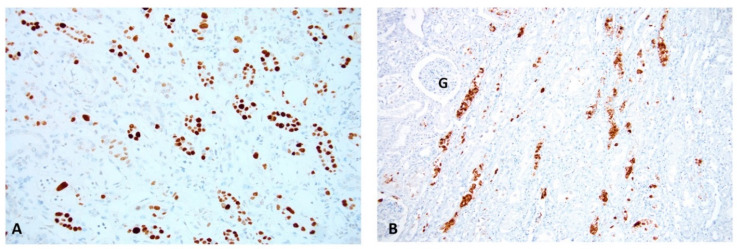
Figure 2.
PyV replication in “definitive” PyVN. Renal Biopsy: (A) An immunohistochemical stain for the SV40-T antigen shows PyV replication with many positive (brown) staining signals in tubular epithelial cell nuclei. This stain targets an early PyV gene product/an intra-nuclear protein associated with PyV replication. (B) An immunohistochemical stain for PyV capsid protein (VP1) demonstrates strong staining (brown) for late PyV gene products. Abundant staining is found not only in nuclei, but also in tubular lumens post release of daughter virions from lysed tubular cells, (compare to Figure 2A and Figure 3). Immunohistochemistry on formalin fixed and paraffin embedded tissue sections, (A) antibody directed against the SV40 T antigen, 200× magnification. (B) antibody directed against the polyomavirus VP1 capsid protein, 100× magnification; (G, glomerulus).

