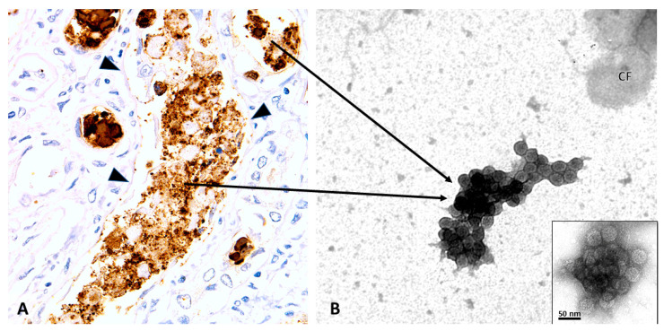
Figure 3.
PyVN, intra tubular viral aggregation and urinary PyV-Haufen. Renal Biopsy (A) and Voided Urine Specimen (B). (A) Immunohistochemistry with an antibody directed against the PyV-VP1 capsid protein showing intra tubular viruses (brown) released into an injured tubule post host cell lysis. Note: denudation of tubular basement membranes. It is here that viruses form dense three-dimensional aggregates, seen as granules of varying sizes by light microscopy (LM). PyV aggregates are subsequently flushed out of the kidney and can be found in the urine as PyV-Haufen by negative staining EM. (B) EM showing characteristic PyV Haufen in a voided urine sample. These PyV Haufen can be easily identified based on the uniform size of the virions and the capsid surface structure (inset). Note the three-dimensional cast-like shape of the PyV Haufen (compare to Figure 4). (A) Immunohistochemistry on formalin fixed and paraffin embedded tissue sections with an antibody directed against PyV-VP1 capsid protein, 400× magnification; (arrowheads, tubular basement membrane; arrows, pointing from the “birthplace” of PyV aggregates to a typical PyV Haufen seen by EM in a voided urine sample in (B). (B) EM with negative staining/uranyl acetate stain, 80,000× magnification, transmission electron microscopy; inset with 120,000× magnification (CF, cell fragment).

