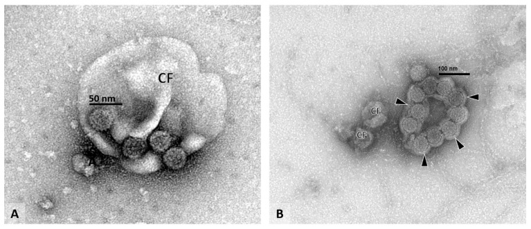
Figure 4.
Voided urine sample with individual virions. Voided urine specimen (A,B), EM: (A) Four individual polyomaviruses can be seen clinging to the surface of a cell fragment (CF). Note the typical viral capsid structure and the uniform size of the viral particles measuring approximately 40 nanometers in diameter. (B) A flat sheet of polyomaviruses can be seen covered by a thin layer of cell membrane material (arrowheads). The characteristic viral capsid structure is visible on each virion. Since tight three-dimensional PyV aggregates are not present, the illustrated findings in (A,B) do not represent PyV-Haufen (compare to Figure 3B). (A) EM with negative staining/uranyl acetate stain, 100,000× magnification, (CF, cell fragment). (B) EM with negative staining/uranyl acetate stain, 80,000× magnification, (CF, cell fragment).

