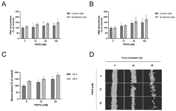
Figure 2.
Extracellular PEPD-dependent proliferation and migration of fibroblasts in a model of closure/scratch assay. (A,B) Control, as well as “scratched” fibroblasts, were treated with PEPD (1–100 nM) for 24 h and 48 h, and proliferation was evaluated using CyQuant Proliferation assay. (C,D) PEPD-stimulated fibroblasts migration was calculated using ImageJ software (https://imagej.nih.gov/ij/) vs. control. PEPD-treated cells were scratched and monitored using an inverted microscope (40× magnification) at 0, 24, and 48 h. Mean values ± SD of three experiments done in replicates are presented. The results are significant at a, b < 0.05, and indicates a vs. control (0 nM of PEPD) of control cells and part C of 24 h incubation, b vs. control (0 nM of PEPD) of scratched cells, and part C of 48 h incubation, respectively. PEPD—prolidase.

