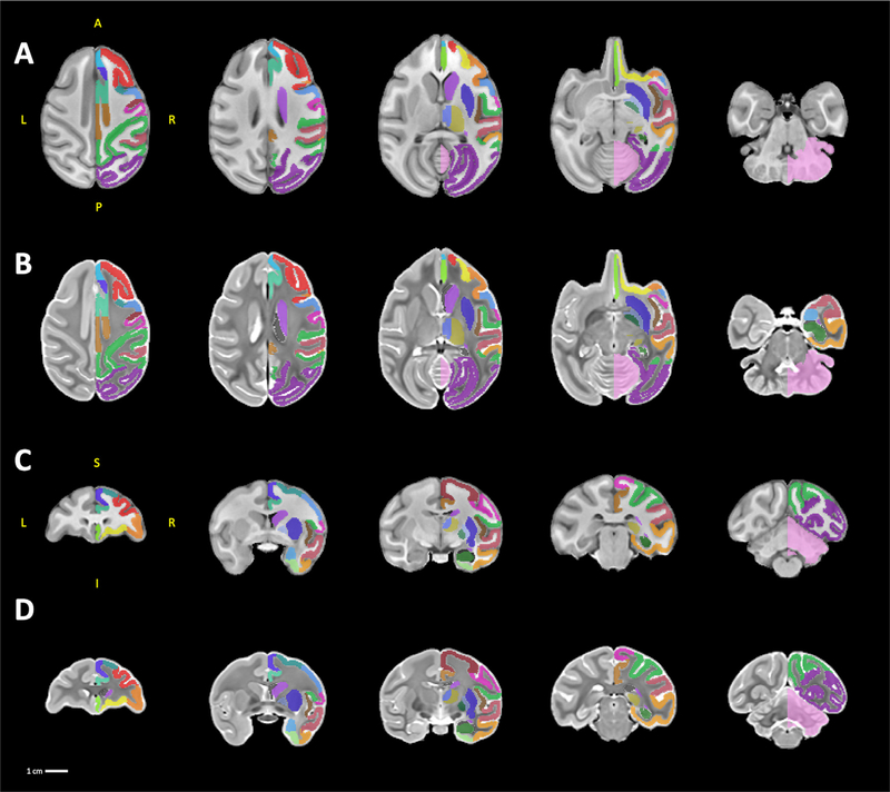
Fig. 2.
sMRI templates. A series of five axial and coronal sections displayed in neurological orientation through the T1 templates (A, C) and the T2 templates (B, D). For ease of viewing, the labelmap overlays are drawn only on the right hemisphere. Abbreviations: A, anterior; I, inferior; L, left; P, posterior; R, right; S, superior.

