Figure 8.

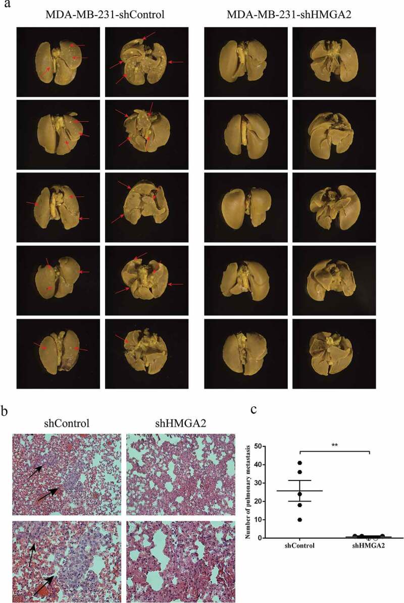
HMGA2 depletion suppresses metastasis in amouse xenograft model
(a): MDA-MB-231 shControl and shHMGA2 cell suspensions containing 2 × 10⁷ cells were injected into the caudal vein. Obvious metastasis was observed in the shControl group, represented by red arrows. (b): HE staining was performed and observed and photographed under the microscope of 200× and 400×, respectively. Obvious abnormal cells were observed in the shControl group, represented by black arrows. (c): the number of metastases in the shControl group was significantly higher than that in the shHMGA2 group. (*P<.05, **P<.01, ***P<.001)
