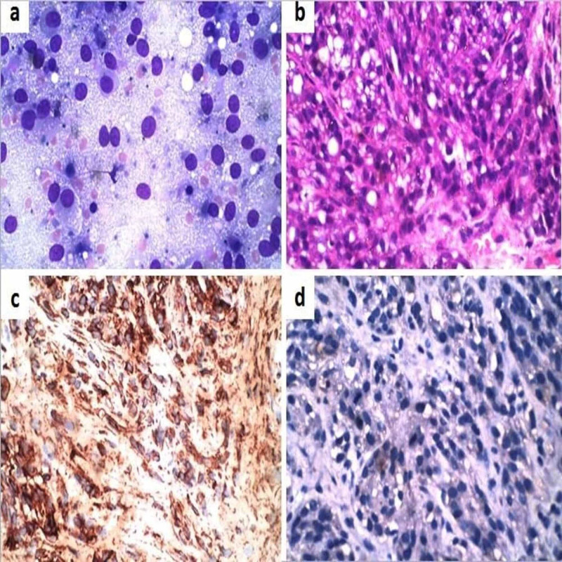
Figure 4. Clear cell sarcoma (a) Smears showed large polygonal cells with abundant wispy cytoplasm, round to oval nuclei, and prominent nucleoli (MGG, 400x); (b) Histopathology showed polygonal to spindle-shaped cells with clear to eosinophilic cytoplasm arranged in fascicles. (H & E, 200x); (c) Tumour cells were positive for HMB 45 (IHC, 200x); (d) Tumour cells negative for cytokeratin (IHC, 200x).
MGG: May-Grunwald Giemsa; H & E: Hematoxylin and eosin; IHC: Immunohistochemistry

