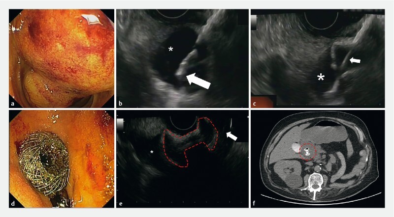
Fig. 1.

Biliary drainage in advanced pancreatic adenocarcinoma: EUS-guided choledocoduodenostomy by LAMS placement. a Inaccessible major papilla due to neoplastic infiltration. b Distal cautery tip of LAMS catheter inside the CBD (asterisk). c LAMS catheter (white arrow) and distal flange fully deployed inside the biliary system (asterisk). d Endoscopic view of proximal flange of the LAMS, perfectly positioned in the duodenal bulb. e EUS evaluation of LAMS profile (dashed red line) with the distal flange in the CBD (asterisk) and proximal one in the duodenum (white arrow). f Computed tomography confirmed the correct position of the LAMS (dashed red circle) 72 hours later.
