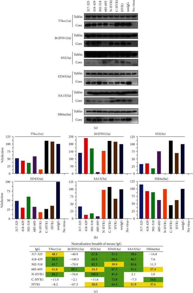
Figure 3.

Neutralization activity of antiserum IgG. (a) IgG (100 μg/mL) induced by each peptide was mixed with 400 FFU of HCVcc for 1 h and added to Huh7.5 cells preseeded in a 24-well plate. Seventy-two hours later, infectivity was evaluated by Western blotting. nmIgG: normal mouse IgG. (b) The density of each band was analyzed with ImageJ software, and the core/Tublin ratio was calculated. The infection of nmIgG was set to 100%, and the relative infection of each group was calculated with GraphPad Prism 8 software. (c) Neutralization breadth of mouse IgG (100 μg/mL). Green, neutralization is >50%; yellow, neutralization is 20%-50%.
