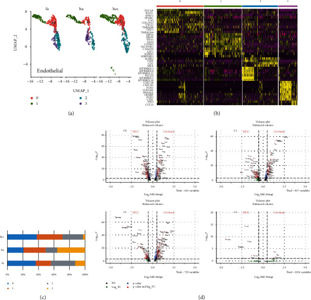
Figure 2.

Subcluster analysis and quantification of the liver endothelial cells. (a) UMAP diagram depicts 4 subtypes of the endothelial cells. (b) Heat map annotated differential genes of 4 endothelial cell subtypes. (c) Bar graphs quantify and compare the proportion of endothelial cell subtypes of healthy, fibrosis, and HCC patients. (d) Volcano plots show the DEGs between fibrosis and HCC in 4 subtypes.
