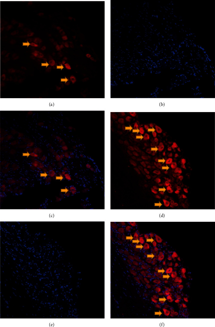
Figure 3.

Distribution of PKH26-labeled SHED in the ipsilateral TG: (a) PKH26-positive cells (red) 24 h after local injection; (b) DAPI-stained cells (blue) 24 h after local injection; (c) the merged image of (a) and (b); (d) PKH26-positive cells (red) 72 h after local injection; (e) DAPI-stained cells (blue) 72 h after local injection; (f) the merged image of (d) and (e).
