
FIGURE 4.
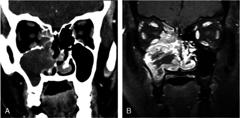
Computed tomography and MRI of 50-year-old woman with pathologically diagnosed SNEC in right maxillary sinus. A, Coronal CT image shows that tumor has moderate enhancement and extending into nasal cavity, ethmoid sinus, and orbit. B, Coronal contrast-enhanced MR image reveals heterogeneous markedly enhanced tumor with necrotic areas.