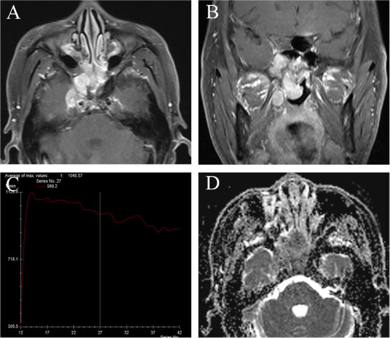
FIGURE 5.

Magnetic resonance imaging of 49-year-old man with SNEC in bilateral sphenoid sinuses. A, Axial enhanced MRI and (b) coronal enhanced MR image reveal the markedly enhanced tumor in sphenoid sinuses. Tumor shows bilateral asymmetry pattern and extending to bilateral nasal choana, right foramen ovale, and cavernous sinus. (C) The TIC examination revealed washout type. D, The ADC map revealed the mean ADC value of 0.724 × 10⁻³ mm2/s.
