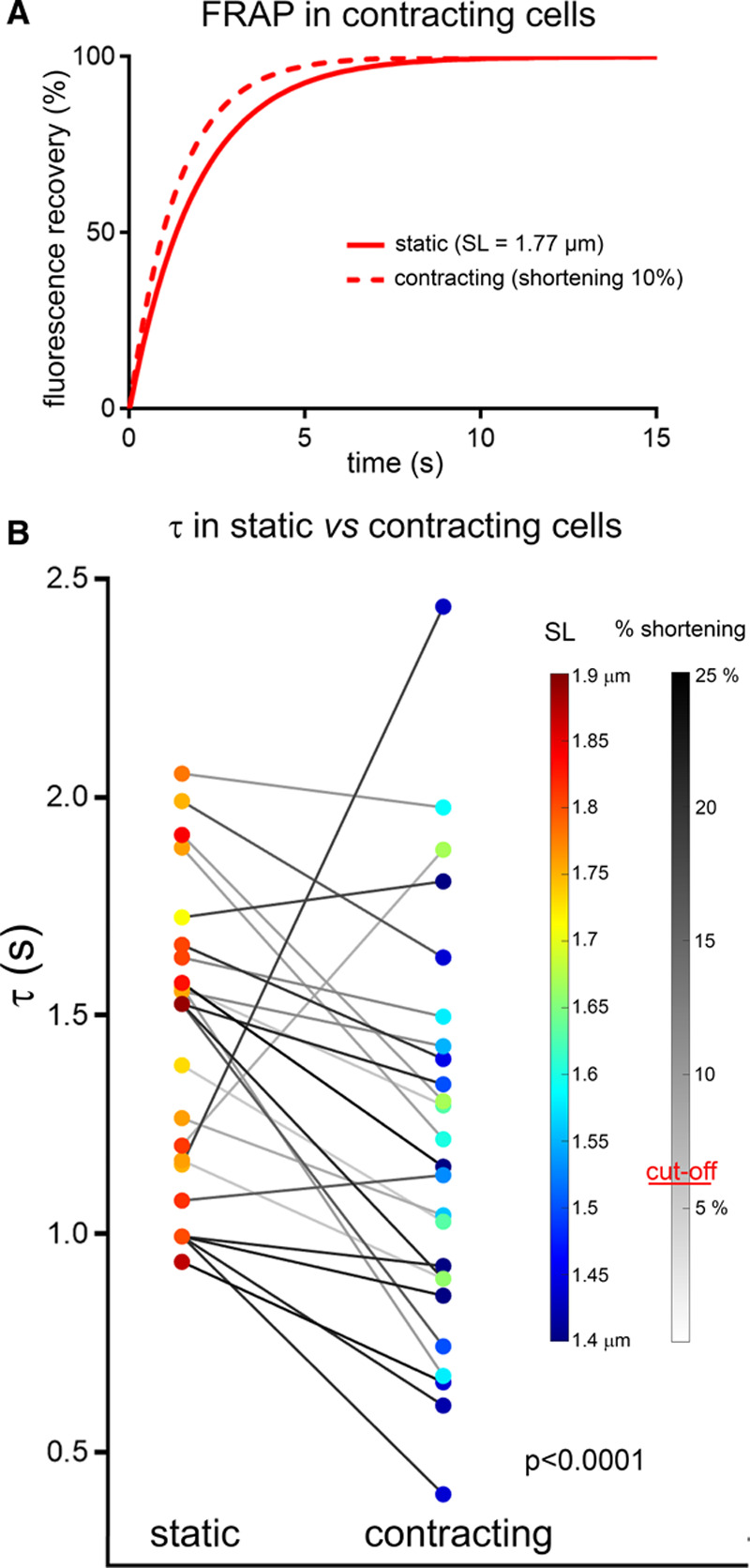
Figure 4.

Apparent speed of transverse tubule diffusion is increased during dynamic contraction and relaxation (live cells). A, Representative fitted fluorescence recovery after photobleaching (FRAP) curves, obtained from one and the same cell at rest and during field stimulation-induced contractions (here at 0.8 Hz). B, Fluorescence recovery times (τ) in cells at rest and during rhythmic contractions (0.5–1.5 Hz, paired observations). Both diastolic and end-systolic sarcomere length (SL) are color-coded, and the extent of percentage shortening during contraction is indicated by the gray-level of connecting lines. Data analyzed using paired T-test, random mixed effect model confirmed a lack of significant effects of heart and cell. N=5 hearts/25 cells.
