Figure 1.
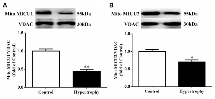
The expressions of mitochondrial MICUs were reduced in Ang-II-induced hypertrophic hearts. (A, B) Western blotting was used to determine the protein expression levels of mitochondrial MICU1 (A) and MICU2 (B) in control and hypertrophic mouse hearts. MICUs, mitochondrial calcium uniporter; MICU1, mitochondrial calcium uptake 1; MICU2, mitochondrial calcium uptake 2. VDAC, voltage-dependent anion channel. VDAC was used as a loading control. Presented values are means ± SEM. N=6-8/group. *P<0.05, **P<0.01 vs. Control.

