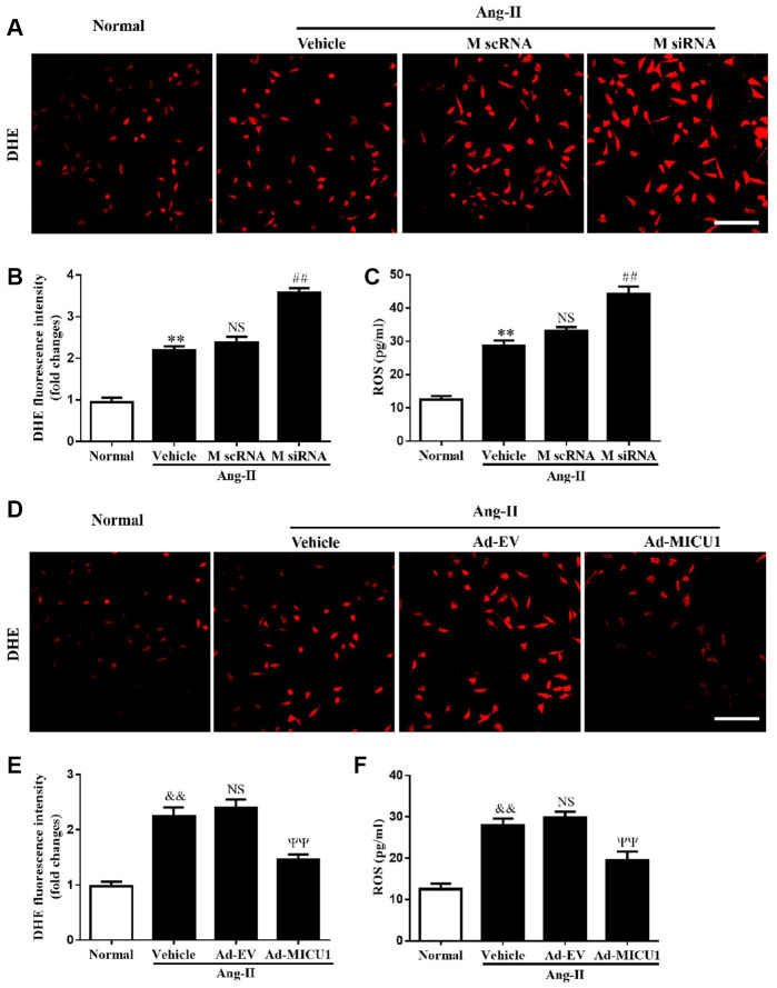
Figure 6.
MICU1 mediated cardiomyocyte hypertrophy by modulating oxidation states. (A, B) The ROS levels in cardiomyocytes treated with Ang-II and MICU1 siRNA were analyzed by DHE staining. Representative confocal microscope images (A) and fluorescence quantitation (B) were presented. Scale bars=10 μm. (C) ROS generation in cardiomyocytes treated with Ang-II and MICU1 siRNA was detected by an ELISA kit. (D, E) The ROS levels in NMVMs treated with Ang-II and Ad-MICU1 were analyzed by DHE staining. Representative confocal microscope images (D) and fluorescence quantitation (E) were presented. Scale bars=10 μm. (F) ROS generation in cardiomyocytes treated with Ang-II and Ad-MICU1 was detected by an ELISA kit. Presented values are means ± SEM. N=6-8/group. **P<0.01 vs. Normal (1); ##P<0.01 vs. M scRNA of Ang-II; &&P<0.01 vs. Normal (2); ΨΨP<0.01 vs. Ad-EV of Ang-II.

