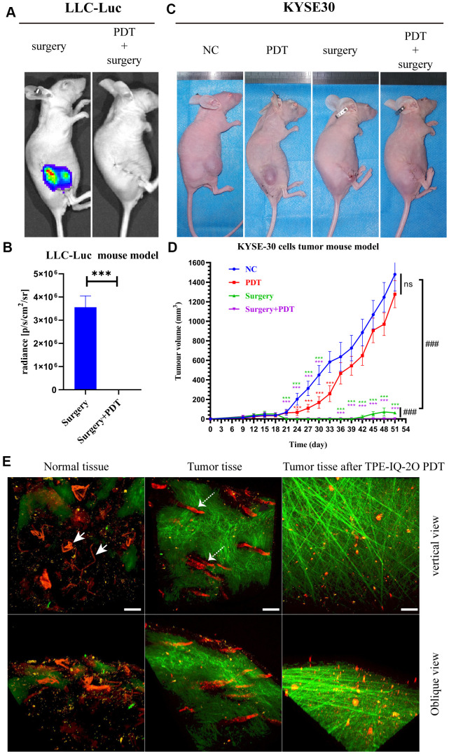
Figure 4.
TPE-IQ-2O PDT combined with surgery can effectively remove the residual tumor focus at the incisal margin and reduce tumor recurrence. (A, B) In vivo bioluminescence imaging and measurement of the LLC-Luc cell tumor mouse model after surgery alone or combined with intraoperative PDT therapy on the 17th day. n=3 mice/group. Data are expressed as the mean±s.d., ***p < 0.001 (C, D) A KYSE-30 cell tumor mouse model and tumor volumes in the control group, PDT alone group, surgery alone group and surgery combined with intraoperative PDT group. n=5 mice/group. Data are expressed as the mean±s.d., ***p < 0.001 vs. control group following the Dunnett or Dunnett’s T3 test, ###p < 0.001 for pairwise comparison on day 51 following Dunnett’s T3 post hoc test. (E) Evaluation of tumor vessels and the stroma after TPE-IQ-2O PDT in the KYSE30 cell subcutaneous tumor model (the white solid arrow shows subcutaneous vessels in normal skin, and the white dashed arrow shows tumor vessels; λex: 1040 nm, λem: 575-610 nm; the green signal represents the stroma, λem: 805 nm; the red signal represents vessels, scale bar=20 μm). (PDT represents the abbreviation of TPE-IQ-2O PDT in this figure).

