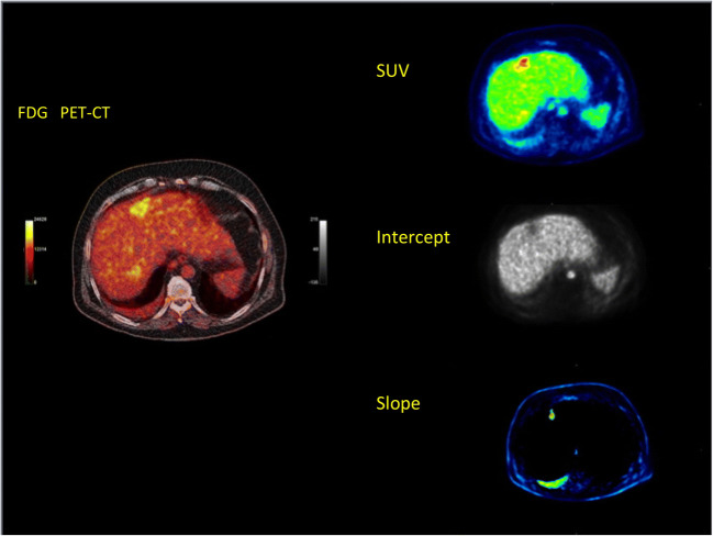
Fig. 5.
Patient with a liver metastasis of rectal cancer following FOLFOX chemotherapy. Fused transversal 18F-FDG PET/CT image (left) demonstrating enhanced uptake at the site of the metastasis 60 min p.i. Transversal SUV image 50–60 min p.i. demonstrates an enhanced uptake (right upper row), while parametric image of the intercept (middle row) shows a decrease in the perfusion-related 18F-FDG uptake, and parametric image of the slope (lower row) demonstrates an enhanced phosphorylation-related 18F-FDG uptake

